Optics labs

The optics (laser) labs in I-FIM utilize spectroscopy and microscopy to study a wide array of heterostructures and layered materials. The spectroscopy measurements involve shining laser excitation on research samples, and detecting their emission/absorption spectra as a function of the excitation energy via high-resolution low-noise spectrometers and cameras. Further, a comprehensive spectral map of the sample is obtained by manipulating the incident laser excitation to different spots on the microscopic samples using piezoelectric positioners with nanometer range. The excitation components include multiple lasers covering the full spectrum from Ultra Violet (UV), through visible to Infra-Red (IR), as well as ultrafast femtosecond lasers to perform time-resolved studies. Broadly, there are two categories of experimental setups for performing optical measurements – (a) the room temperature apparatus to allow ambient characterization and (b) low temperature configurations which provide the additional measurement knobs of temperature and magnetic field.  

List No. 

Key specifications for existing equipment 

Picture 
A.  Spectrometers 

Princeton spectrograph with 750 mm focal length and scanning monochromator that features an astigmatism-corrected optical design, a mechanical scanning range of 0–1500 nm, as well as resolution of >0.05 nm. Equipped with avalanche photodiodes, CCD’s and InGaAs detectors to facilitate different measurements such as Raman, photoluminescence, fluorescence, transmission and absorption. 

Spectrograph 1 

    • With 3 gratings from 300g/mm BLAZE (500nm) to 1800 g/mm BLAZE (500nm) 
    • Detector (x1) Liquid nitrogen cooled CCD camera with UV coating (Peak QE from UV to NIR) 

Spectrograph 2 

    • With 3 gratings from 1200g/mm BLAZE (750nm) to 150 g/mm BLAZE (1.6µm) 
    • Detector (x1) Liquid nitrogen cooled CCD camera (Peak QE from UV to NIR) 
    • Detector (x1) Liquid nitrogen cooled InGaAs photodiode array (Spectral range 0-2.2 μm) 

Spectrograph 3 

    • With 3 gratings from 150g/mm BLAZE (500nm) to 1800 g/mm BLAZE (500nm) 
    • Detector (x1) Liquid nitrogen cooled CCD camera with UV coating (Peak QE from UV to NIR) 

Spectrograph 4 

    • With 3 gratings from 1200g/mm BLAZE (750nm) to 150 g/mm BLAZE (1.6µm) 
    • Detector (x1) Liquid nitrogen cooled CCD camera (Peak QE from UV to NIR) 
    • Detector (x1) Liquid nitrogen cooled InGaAs photodiode array (Spectral range 900-1700 nm) 
B. Cryostat 1 

Cryogen free closed-loop system with a cold objective and low temperature scanning probe microscope (SPM). Extremely useful to produce spectral maps within short duration.  

    • Allows quick sample exchange (top loading insert)  
    • No magnet 
    • Base temperature <4 Kelvin  
    • Cooldown time <3 hours 
    • Supports optical fiber coupled or free beam excitation 
    • Piezo based nanopositioners for closed loop XYZ positioning  
    • Supports DC transport with 8 sample contacts (max)   
C. Cryostat 2 

Cryogen free closed-loop system with 2 inserts – 1) a scanning confocal microscope (CFM) with cold objective, 2) an insert to perform low-temperature atomic force microscopy (AFM) and magnetic force microscopy (MFM).  
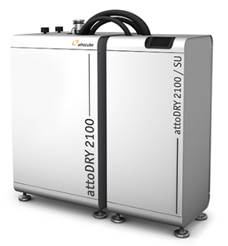
    • Allows quick sample exchange (top loading insert)  
    • Superconducting Magnet ±9 Tesla (vertical) 
    • Base temperature 1.6 Kelvin (needle valve controlled closed loop Helium circulation)  
    • Cooldown time <5 hours 
    • Piezo based nanopositioners for closed loop XYZ positioning  
    • CFM insert supports both Voight and Faraday geometry for sample mounting 
    • CFM supports optical fiber coupled or free beam excitation 
    • AFM/MFM insert Signal-to-Noise ratio at LT > 20:1 
D. Cryostat 3 

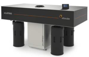
Low-vibration cryocooler integrated into an optical table to allow flexible switching between different optical excitation and detection paths, via easy manipulation of the optomechanical elements. 

    • Equipped with 2 separate easily mountable vacuum shrouds – 1 with a cold objective inside the shroud and 1 with a room-temperature objective placed outside on the optical table. 
    • Base temperature <4 Kelvin  
    • No magnet 
    • Cooldown time <6 hours 
E. Cryostat 4:

Cryogen free closed-loop system with 3 inserts – 1) a scanning confocal microscope (CFM) with cold objective, 2) an insert to perform low-temperature atomic force microscopy (AFM) and magnetic force microscopy (MFM) 3) an insert with a built in Diamond Anvil Cell (DAC) for high pressure (>10 (GPa) microscopy

  • Allows quick sample exchange (top loading insert)
  • Superconducting Vector Magnet ±9 Tesla (z), ±3 Tesla (x)
  • Base temperature 1.6 Kelvin (needle valve controlled closed loop Helium circulation)
  • Cooldown time <5 hours
  • Piezo based nanopositioners for closed loop XYZ positioning
  • CFM insert supports both Voight and Faraday geometry for sample mounting
  • CFM supports optical fiber coupled or free beam excitation
  • AFM/MFM insert Signal-to-Noise ratio at LT > 20:1


F. Lasers 

List of existing lasers to cover the wide spectrum of materials being studied 

    • Argon (450-530 nm) 
    • HeNe (543, 633 nm) 
    • Supercontinuum (350-2600 nm) 
    • Er Fiber laser (785 nm) 
    • Vanadate (532 nm) 
    • Sapphire (700-1000 nm) 
    • MOFA (266, 355, 532 nm) 
    • Fiber (780/1560 nm) 
    • OPO (690-2300 nm) 
    • Diode (404, 532.1, 784.5, 405, 488, 532, 660, 730, 1064, 375, 515, 534, 640, 785, 980, 514.4 nm)